Hip Muscles Diagram : Why Hip Flexors Are Tight And Why Your Hips Pop Sparta Science : The iliacus muscle, the psoas major muscle, and the psoas minor muscle.. A hip strain occurs when one of the muscles supporting the hip joint is stretched beyond its limit or torn. They can be divided into three main groups: You can pull your toes up at the exact same time to add another measurement to the stretch. It allows for complete rotations of the hip and is also. Hip pain, especially while it rotates in certain directions.
Beginner hip flexor muscle anatomy. Scroll down to see the muscle names that go with these letters. They allow you to move your leg or knee up towards your torso, as well as to bend your torso forward at the hip. Labral tears are difficult to diagnose, partially because of the many muscles and other structures that are near the hip joint. A sprain is a torn or overstretched ligament while a strain is a torn or overstretched tendon or.
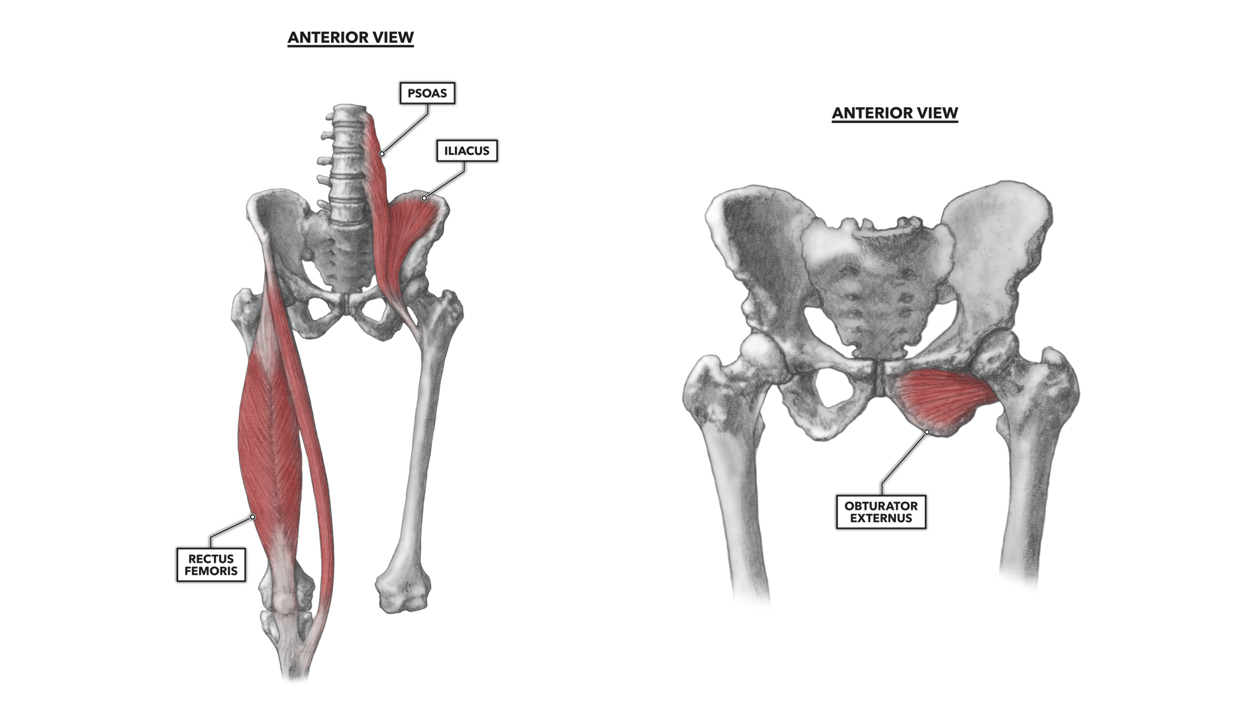
Tendinitis And Bursitis Treatment Cincinnati Tendinitis Dayton Oh from www.beaconortho.com The iliopsoas actually consists of two muscles: Browse 4,822 hip anatomy stock photos and images available, or search for hip replacement or knee anatomy to find more great stock photos and pictures. A hip strain occurs when one of the muscles supporting the hip joint is stretched beyond its limit or torn. In human anatomy, the muscles of the hip joint are those muscles that cause movement in the hip.most modern anatomists define 17 of these muscles, although some additional muscles may sometimes be considered. The four muscle of the quadriceps all extend the lower leg, and the rectus femoris additionally can flex the thigh at the hip. The muscles work together to enable movement and keep the hip in alignment. The quick answer to this question is the muscles of the lower back are the multifidus, longissimus, spinalis, and quadratus lumborum. Use acronyms to help you.
They can be divided into three main groups:
It allows for complete rotations of the hip and is also. Utilizing your hands, gently push up until your spine is straight. These are often divided into four groups according to their orientation around the hip joint: A hip strain occurs when one of the muscles supporting the hip joint is stretched beyond its limit or torn. The psoas major, the longer of the two muscles, originates on the lumbar vertebrae and attaches to the femur. The many muscles of the hip provide movement, strength, and stability to the hip joint and the bones of the hip and thigh. They allow you to move your leg or knee up towards your torso, as well as to bend your torso forward at the hip. When you flex your hip, you move the leg forward. Muscles play an important role in the. The iliacus originates on the pelvic crest and attaches on the femur. Extension, flexion, adduction, and abduction. Hip diagram slide your left leg back until the top of your thigh rests on the ground. The quick answer to this question is the muscles of the lower back are the multifidus, longissimus, spinalis, and quadratus lumborum.
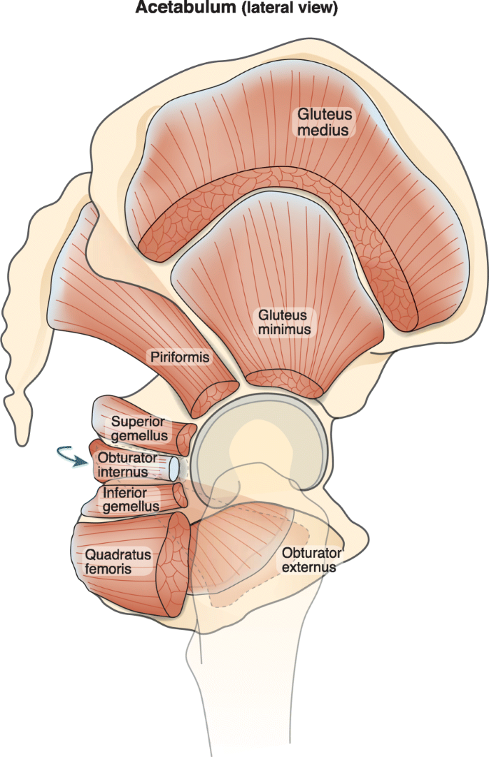
Hip diagram slide your left leg back until the top of your thigh rests on the ground. Aaa i gg pp r s t. Iliopsoas muscle (musculus iliopsoas) iliopsoas is a large compound muscle of the inner hip composed of the iliacus and psoas major muscle side from the iliopsoas, other muscles of the inner hip include the psoas minor, obturator externus, obturator internus, superior gemellus, inferior gemellus, piriformis and. Smartdraw includes 1000s of professional healthcare and anatomy chart templates that you can modify and make your own. In simple terms, these muscles rotate the front of your pelvis and the front of your thighs toward each other.
Crossfit Hip Musculature Part 1 Anterior Muscles from www.crossfit.com The four groups are the anterior group, the posterior group, adductor group, and finally the abductor group. Hip diagram slide your left leg back until the top of your thigh rests on the ground. The iliopsoas muscle is a major mover of your hip joint. A severe strain can limit your ability to move your hip. In human anatomy, the muscles of the hip joint are those muscles that cause movement in the hip.most modern anatomists define 17 of these muscles, although some additional muscles may sometimes be considered. Smartdraw includes 1000s of professional healthcare and anatomy chart templates that you can modify and make your own. The muscles in the hip are responsible for the movement of the hip and, by proxy, the leg. These muscles work together to flex your hip and to stabilize your hip and lower back during activities such as walking, running, and rising from a chair.
The quick answer to this question is the muscles of the lower back are the multifidus, longissimus, spinalis, and quadratus lumborum.
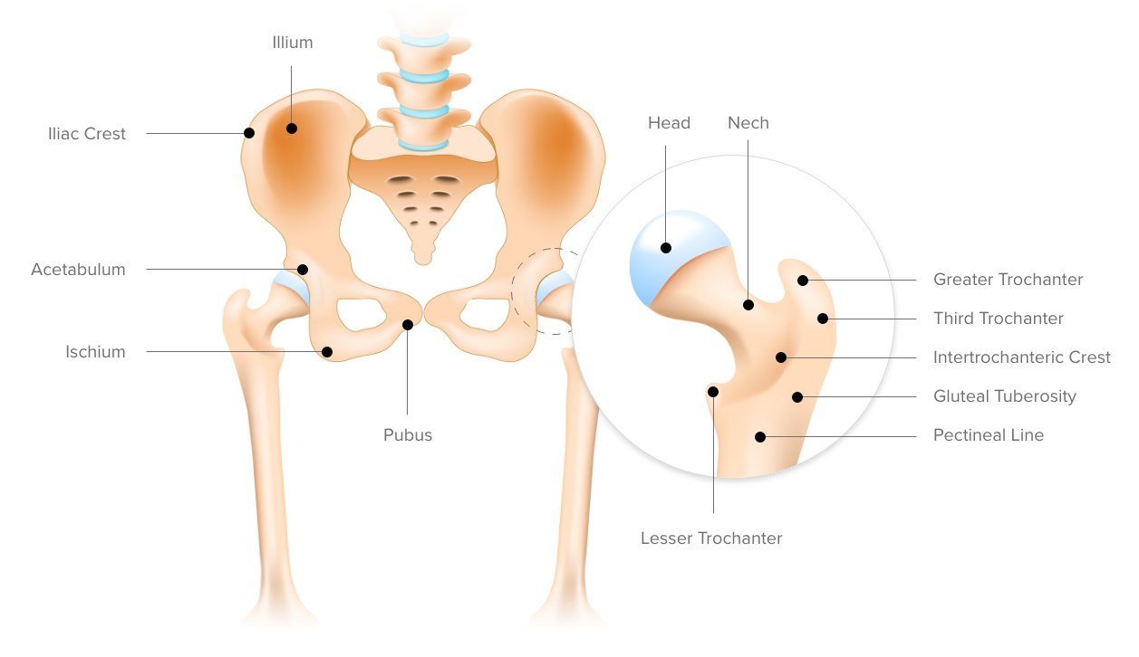
The quick answer to this question is the muscles of the lower back are the multifidus, longissimus, spinalis, and quadratus lumborum. The hip muscle diagram below shows a number of the muscles we will be discussing in the next sections. The four muscle of the quadriceps all extend the lower leg, and the rectus femoris additionally can flex the thigh at the hip. A feeling or sound of clicking or locking when your hip is in motion. They can be divided into three main groups: Hip diagram slide your left leg back until the top of your thigh rests on the ground. The hip flexors can be found connecting the top of the femur, which is the largest bone in the body, to the lower back, hips, and groin. You can strain or tear your hip flexor muscles through sudden movements or falls. The psoas major, the longer of the two muscles, originates on the lumbar vertebrae and attaches to the femur. A sprain is a torn or overstretched ligament while a strain is a torn or overstretched tendon or. More commonly, our hips flex to a 90° angle when we sit. This article will introduce the muscles in each group and touch on their origin, insertion, function, and innervation. Iliopsoas muscle (musculus iliopsoas) iliopsoas is a large compound muscle of the inner hip composed of the iliacus and psoas major muscle side from the iliopsoas, other muscles of the inner hip include the psoas minor, obturator externus, obturator internus, superior gemellus, inferior gemellus, piriformis and.
The four groups are the anterior group, the posterior group, adductor group, and finally the abductor group. The iliacus muscle, the psoas major muscle, and the psoas minor muscle. A sprain is a torn or overstretched ligament while a strain is a torn or overstretched tendon or. Gluteal muscles, located on the back of the hip (buttocks) · the adductor muscles on the inner thigh bring. The anterior muscle group features muscles.
Architecture Of The Short External Rotator Muscles Of The Hip Bmc Musculoskeletal Disorders Full Text from media.springernature.com The iliacus muscle, the psoas major muscle, and the psoas minor muscle. These are often divided into four groups according to their orientation around the hip joint: More commonly, our hips flex to a 90° angle when we sit. Large ligaments, tendons, and muscles around the hip joint hold the bones (ball and socket) in place and keep it from dislocating. These muscles connect the front of your pelvis to the front of your thigh (the psoas also connects to your spine). Scroll down to see the muscle names that go with these letters. Anyone can experience a hip strain just doing everyday tasks, but strains most often. If you're just starting your anatomy journey, work on remembering the names of all 11 hip flexor muscles.
Large ligaments, tendons, and muscles around the hip joint hold the bones (ball and socket) in place and keep it from dislocating.
Push down carefully, leaning just as far as you can without overextending your hips. The rectus femoris is also a hip flexor. The diagram shows the posterior (rear) view of the buttock. Here are the letters to work with: Use acronyms to help you. Beginner hip flexor muscle anatomy. Best program for drawing diagrams on windows and mac. The iliacus muscle, the psoas major muscle, and the psoas minor muscle. Utilizing your hands, gently push up until your spine is straight. Large ligaments, tendons, and muscles around the hip joint hold the bones (ball and socket) in place and keep it from dislocating. Ligaments, tendons, and muscles play an important role in the function of the hip. The iliacus originates on the pelvic crest and attaches on the femur. Smartdraw includes 1000s of professional healthcare and anatomy chart templates that you can modify and make your own.